The Eyes Have It!
Recorded On: 10/15/2020
- Registration Closed
Eyes are notoriously a difficult tissue to demonstrate histologically, non-human primate (NHP) eyes being especially challenging. There are standards that need to be followed for all steps of the histological processes in order to achieve a readable slide for the pathologist, without doing so can result in damage to the tissue, poor infiltration, difficulty sectioning, and artifacts that obscure areas of interest on the slide. Within the NHP eye there are several specialized structures for high-acuity vision that can require pathological evaluation, in particular the macula and the fovea. These specialized structures translate to the human eye as well, making their importance in the pharmaceutical industry and clinical setting all the more critical. The basic structures of the eye need special considerations in regards to fixation and handling, but to achieve acceptable morphology of the macula and fovea specific guidelines must be followed when trimming, embedding and cutting. Collection of the eye requires a delicate hand and refined technique to ensure that important landmarks are maintained as well as cellular and structural integrity. Eyes are fixed whole to maintain retinal attachment. Orientation of the left and right eye needs to be maintained along with muscular landmarks to ensure that grossing is done correctly for the macula and fovea to be demonstrated. There are several fixatives that can be used, each with their own benefits and drawbacks; choosing the most beneficial one can be challenging. Even with diligent collection, fixation and processing there can still be difficulties embedding and sectioning the eye. The aim of this presentation is to guide histotechnicians and technologists through the process and potential troubleshooting required for the collection, fixation choice, processing cycles, embedding and microtomy of the NHP eye to achieve a quality specimen that produces a quality slide fit for pathological evaluation. The importance of various eye structures for the pharmaceutical industry and clinical purposes will also be covered as well. All procedures performed on these animals were in accordance with the regulations and established guidelines and were reviewed and approved by an Institutional Animal Care and Use Committee or through an ethical review process.
CEUs: This histology course is worth 1 continuing education credit. Course is available for 365 days from date of purchase.

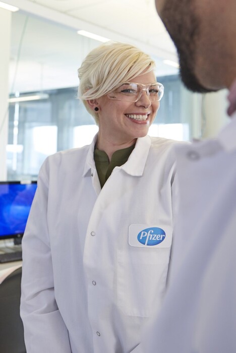
Kelly Voight, HT(ASCP)
Histotech
Pfizer
Currently, I am responsible for performing all aspects of tissue collection through slide review. I necropsy, trim, embed, cut, stain, and QC the tissues for pathologic evaluation. In addition I am a trainer in histology tasks of trim (grossing), embed and microtomy as well as a necropsy trainer. I also work on methods development in our lab and am a liaison for our environmental health surveillance team. I have recently been made a training lead in the lab and am responsible for scheduling trainings as well as the daily tasks. Before working at Pfizer Inc. I worked for the Hartford Healthcare Network at Backus Hospital in the pathology department. I was responsible for embedding and cutting patient tissue, immunohistochemistry staining, and special stains as well as lab equipment maintenance
